As the expert in drug therapy, you need to know how different drugs exert their effects on the electrical system of the heart.
Pharmacist review of the ECG is relatively straight forward compared to other professions. We should be able to identify the rhythm, look for signs of drug toxicity, and calculate a QTc. We don’t need to worry about the different leads, or which lead is looking at which side of the heart, or any of the other complicated nuances other providers need to know.
In this episode I’ll review the ECG components, propose a step-wise approach to identifying the rhythm, and give examples for the following pharmacy related ECG abnormalities:
- Hyperkalemia
- ST-segment elevation MI
- Prolonged QTc interval
- ECG signs of drug toxicity
- Beta blockers
- Digoxin
- Tricyclic antidepressants
- Antiarrythmics
- Dysrhythmias
- Torsades
- Ventricular Tachycardia / Fibrillation
- Atrial Fibrillation
- Heart block
Review of ECG components
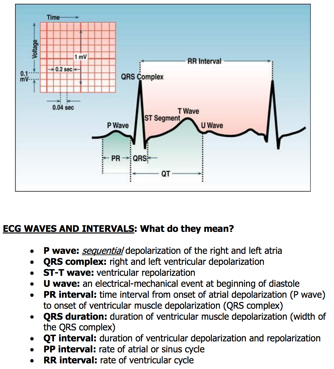
P wave – represents atrial depolarization
QRS complex – represents ventricular depolarization
T wave – represents ventricular repolarization
PR interval – represents time for the impulse to travel from the SA node through the AV node to the ventricles
QT interval – represents the time for ventricular depolarization and repolarization to occur (called the QTc interval when corrected for heart rate)
R-R interval – represents the time between beats and is used to calculate the heart rate
Image courtesy of ecg.utah.edu
Stepwise process to interpreting an ECG
There are many different ways to approach ECG interpretation. Whether you use my approach or another, be sure to use the same process each time.
1. Is there a P wave before each QRS complex? This tells you if the rhythm originates in the SA node.
2. Is the R-R interval regular?
3. Is the rate below 60 or above 100?
4. Is the PR interval normal? It should be less than 5 small boxes on the ECG.
5. Is the QRS complex normal? It should be narrow, 2-3 small boxes on the ECG.
6. Is the QT less than half the R-R interval? If not, the QTc is likely prolonged.
7. Does each QRS complex look the same? If not, this could indicate PVCs or PACs.
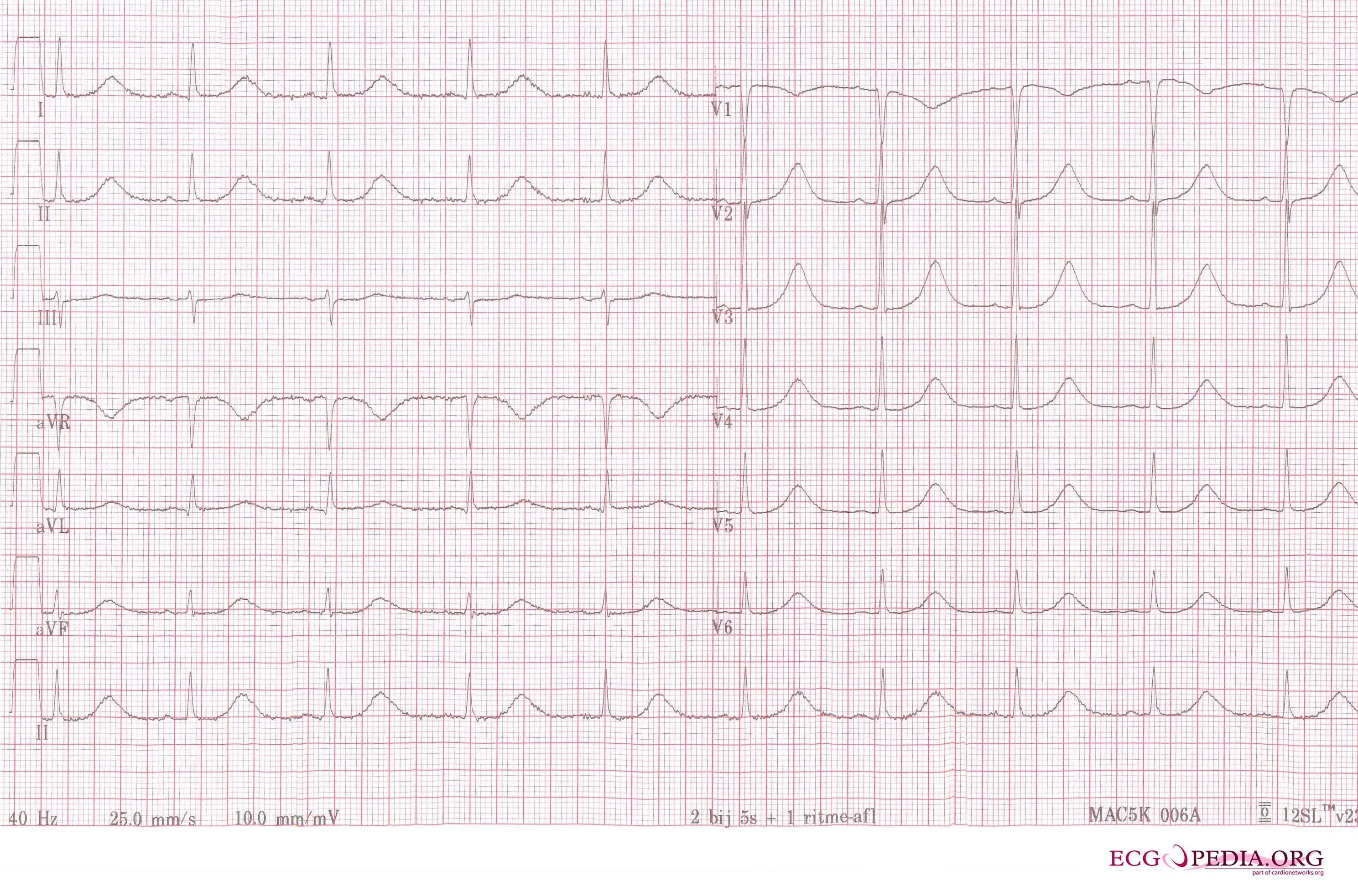
Hyperkalemia
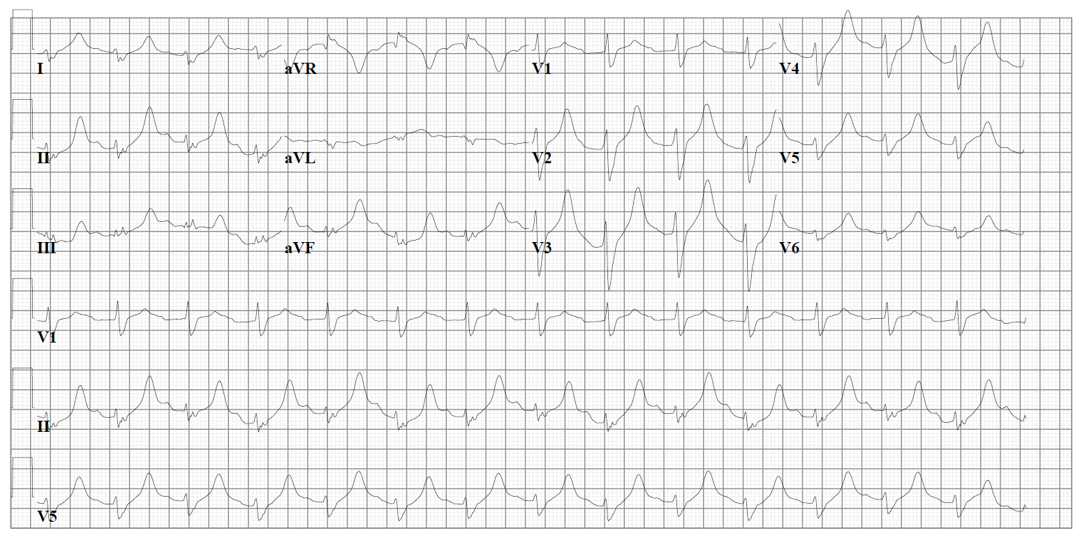
Look for flat or missing P waves, peaked T waves, and wide QRS interval.
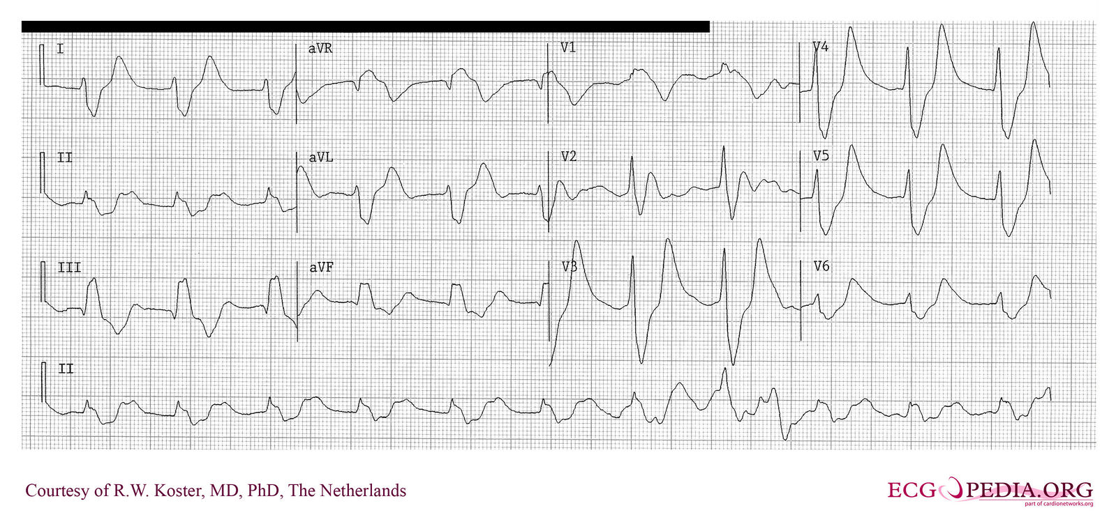
ST segment elevation MI
Look for a markedly elevated ST segment, that appears like a “tombstone”. Diagnosis of acute MI is far more complicated, but I find it valuable to know this basic identification. The ST segment elevation indicates a complete blockage to a coronary artery. The example below is an acute MI with proximal LAD occlusion.
Prolonged QTc interval
In the example below the QTc is about 640 msec (normal is around 450 msec or less). Look how the QT interval is more than half the R-R interval – this is my visual shortcut to determining if I should look harder at how prolonged the QTc is.
Beta-blocker toxicity
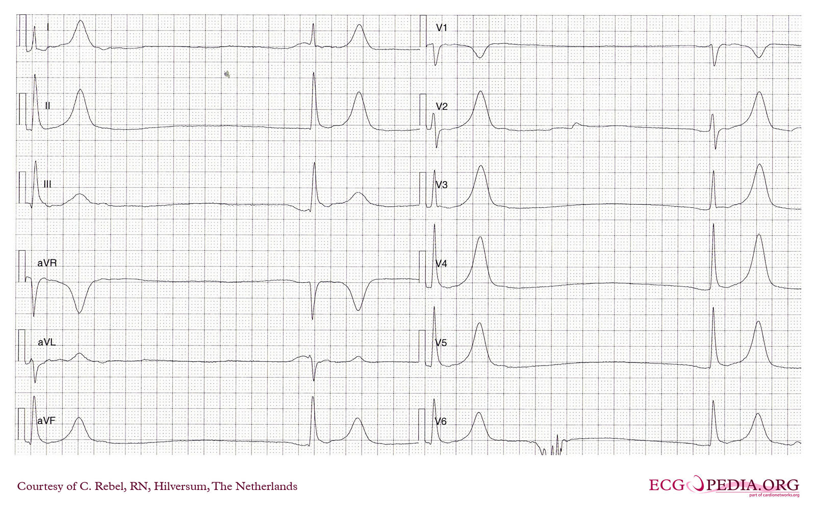
Look for severe bradycardia with QRS widening. The example below is a patient with atenolol toxicity.
Digoxin effect / toxicity
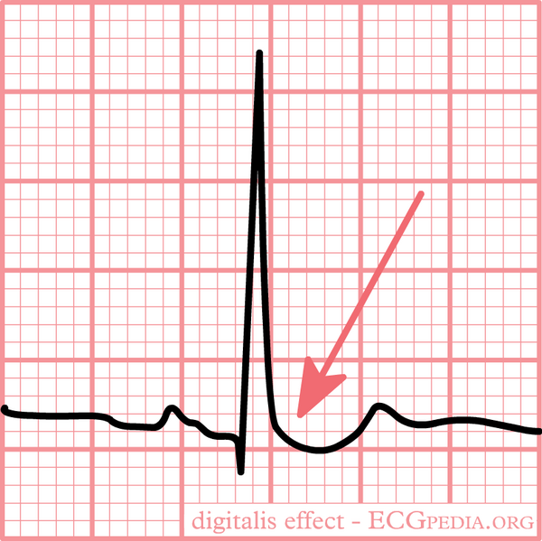
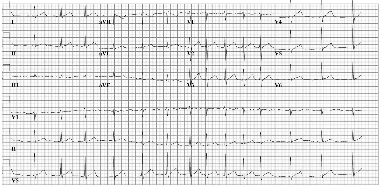
Digoxin causes a funny “scooped” looking ST depression which you can see in the image below. This does not indicate toxicity – just that the patient is taking digoxin.
Digoxin toxicity can cause any number of atrial and ventricular brady and tachyarrthymias. Click here for more details.
Tricyclic antidepressant toxicity
Look for a wide QRS and prolonged QTc. This is caused by sodium channel blockade and can quickly become life-threatening. Push IV sodium bicarb until the QRS narrows then hang a bicarb drip!
Antiarrhythmic toxicity
For antiarrthymic toxicity, look for abnormalities in ventricular conduction (long QTc, wide QRS, etc…).
The example below is of sotalol toxicity. There is a prolonged QTc and a premature ventricular beat.
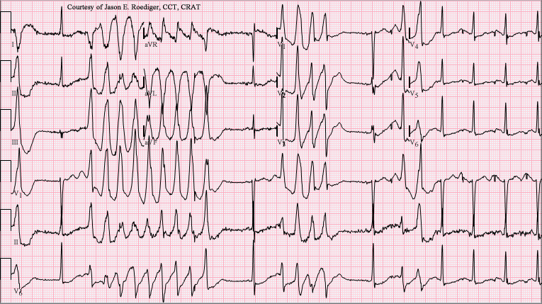
Torsades
According to ecgpedia.org:
During Torsades de Pointes the ventricles depolarize in a circular fashion resulting in QRS complexes with a continuously turning heart axis around the baseline (hence the name Torsades de Pointes).
Below is a 12-lead ECG of Torsades de Pointes (TdP) in a 56-year-old white female with a potassium of 2.4 mmol/L and a magnesium of 1.6 mg/dL.
Ventricular tachycardia / fibrillation
For VTach, look for only ventricular beats (first example below). This is a medical emergency, and can quickly develop into a pulseless rhythm.
VFib (second example below) is a chaotic depolarization of the ventricles and should be treated by immediate CPR and defibrillation. If you encounter VFib in a conscious patient – check for problems with the ECG leads first!
Atrial fibrillation
Look for absence of P waves and irregular R-R intervals – now it is easy to spot AFib!
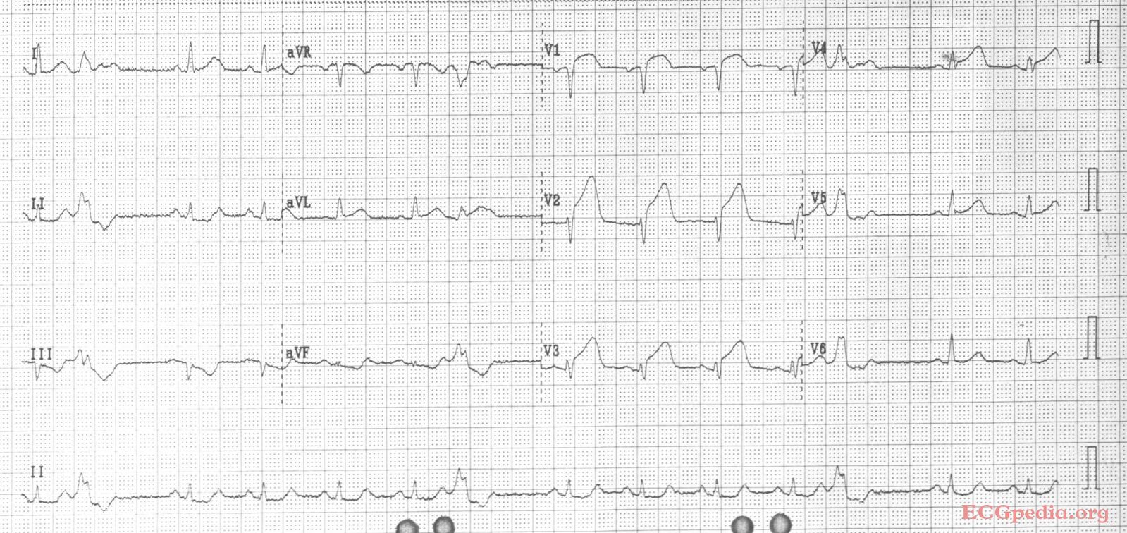
Heart block
There are many types of AV block and I encourage you to review them in the resources provided at the end of this post.
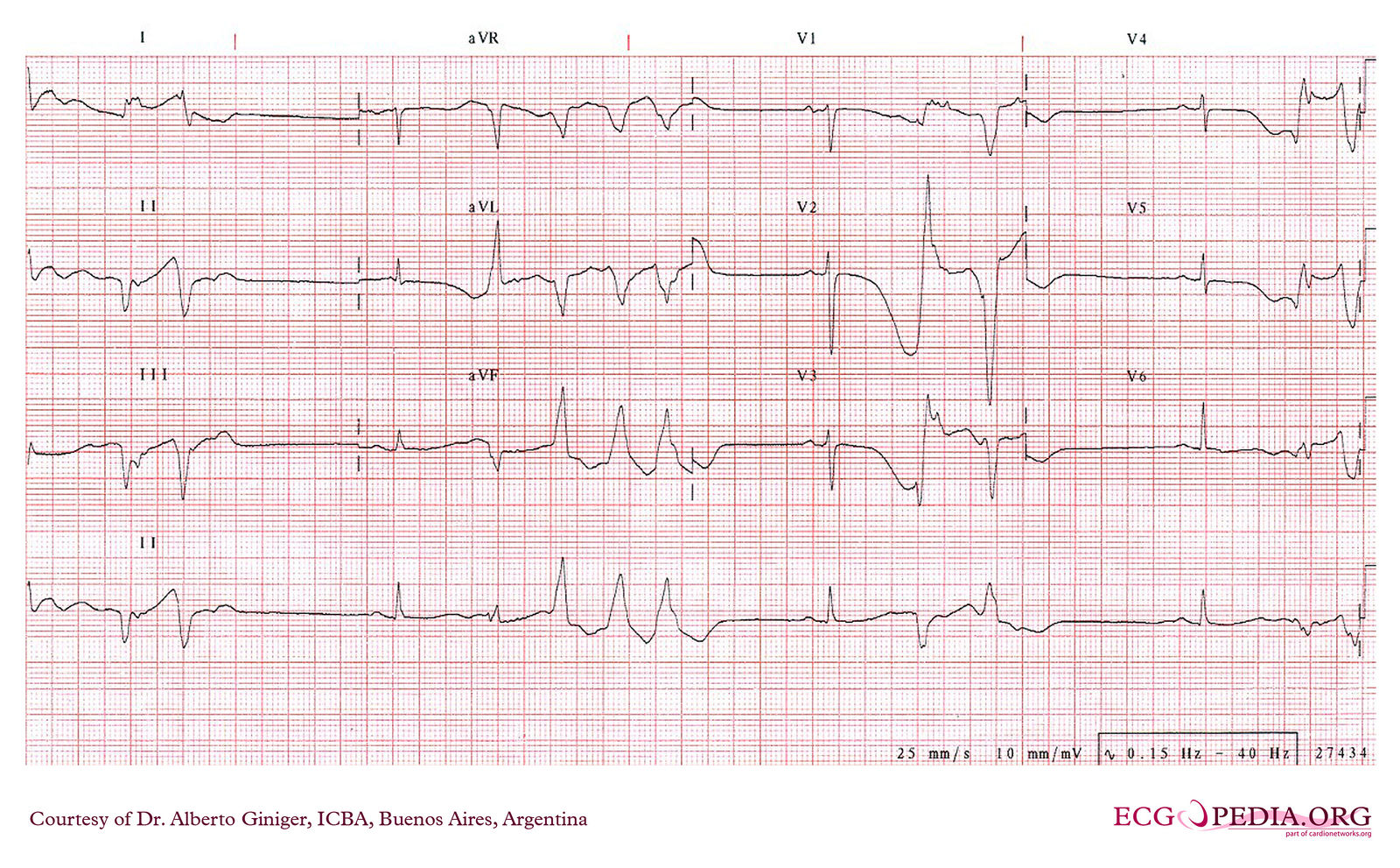
Below are two examples of 3rd degree heart block, which is considered life threatening. The second example was induced by adenosine and is short lasting.
Look for no temporal relationship between the P waves and the R waves. The atria and the ventricles are not in sync due to the complete block of electrical activity between them.
My favorite ECG resources
I encourage you to check out these resources! I developed my ability to look at ECG from a pharmacist’s point of view through self study of these resources and querying my hospital cardiologists to help me make sense of it all. I hope you do the same!
ecgpedia.org (where all of the ECG images for this post came from)
EKG and Rhythm Interpretation 101
Introduction to ECG Interpretation
If you like this post, check out my book – A Pharmacist’s Guide to Inpatient Medical Emergencies: How to respond to code blue, rapid response calls, and other medical emergencies.




Best intro ever!! Love, Aunt Carrie 🙂
Great podcast! This has been helpful in preparing me for my ICU rotation. Could repost the pictures of the ECG. They are no longer showing.
could you*
Thank you for letting me know! They should be all set now.